Abstract
Background: Management strategies for post-Ascyrus Medical Dissection Stent (AMDS) dilation remain poorly described, and surgical approaches are lacking for these complex reoperations.
Case summary: We present a 59-year-old female patient who previously underwent emergency Acute Type A Aortic Dissection (ATAAD) repair with ascending aorta and hemiarch replacement combined with AMDS implantation. Four years later, she developed progressive aortic arch dilation up to 58 mm secondary to a distal anastomosis new entry tear and residual arch vessel dissection.
Surgical details: A hybrid arch replacement strategy with supra-aortic debranching and antegrade deployment of a zone 0 hybrid frozen elephant trunk deployed within the AMDS device
Take-home message: Our strategy illustrates a safe and reliable option for complex reoperation after AMDS implantation without the necessity to explant the device.
Keywords
Dissection; Failure; Aorta.
INTRODUCTION
The Ascyrus Medical Dissection Stent (AMDS) is a hybrid uncovered stent that is indicated for acute type A aortic dissection (ATAAD) repair n to promote false lumen obliteration, true lumen expansion and positive aortic remodeling [1]. Recent literature raised concerns regarding its long-term efficacy. Aortic diameter progression seemed to be a recurrent complication which poses unique challenges for surgical reintervention [2]. Removal of the AMDS is associated with a high, almost prohibitive, risk of damaging the aortic wall with difficult reconstruction of aortic continuity [3]. Description of other surgical techniques, without device removal are required for managing these complex cases of aortic dilatation post-AMDS.
CASE PRESENTATION
We present a 59-year-old female patient treated for an acute De Bakey type 1 aortic dissection four years earlier. She presented with a right upper limb, right lower limb and right carotid artery malperfusion. She was neurologically intact with reduced sensation and strength in both legs. Femoral pulses were absent. She underwent emergency ATAAD repair at our institution. After right axillary cannulation, the ascending aorta and hemiarch were replaced under circulatory arrest. An AMDS was positioned in zone 0 extending into the descending thoracic aorta (Figure 1A). There was no postoperative complication, and she fully recovered from her malperfusion syndrome. She was discharged home on postoperative day 11. In the first year following ATAAD repair, follow-up CT imaging showed no change in the aortic arch and descending aorta diameter. On year 2, a mild enlargement of 3 mm was observed in mid arch, with stable false lumen size. On year 3, the maximum aortic arch diameter had reached 48 mm. On year 4, due to the residual dissection secondary to a distal anastomosis new entry (DANE) tear, and aortic arch dilation up to 58 mm, surgical intervention was indicated. The patient initially underwent a left carotid-subclavian bypass. A few days later, after redo sternotomy, an ascending aorta and full arch replacement was performed under circulatory arrest. A hybrid frozen elephant trunk (COOK FET graft, Bloomington, In) was placed in zone 0, covering the initial distal anastomosis and three head vessels.
SURGICAL DETAILS
Two days following a left carotid-subclavian bypass (Figure 1B), patient underwent redo sternotomy, right atrial venous cannulation and aortic canulation in the ascending aortic graft. The innominate artery and left carotid were debranched and anastomosed on a Y graft where cerebral perfusion could be initiated independently to the systemic cooling (arch first technique) (Figure 1C). A soft guidewire was brought to the ascending aorta through the left femoral artery. Once the patient was cooled to 26 degrees Celsius, circulatory arrest was initiated and the ascending aorta graft was opened in an almost circular fashion leaving only a rim of none transected graft posteriorly.
Under circulatory arrest, over the soft wire, a Cook hybrid prosthesis (26/26/170 mm) was deployed in an antegrade fashion (Figure 1D).The COOK graft has a distal stented portion with a proximal unstented free polyester portion allowing for easy suturing. This way, the ascending aorta, the arch tear (DANE), and proximal descending aorta were excluded. Using 3-0 prolene, an anastomosis was performed incorporating the endovascular prosthesis and the the trimmed polyester of the COOK graft hence closing the previously transected dacron graft incorporating the free polyester graft of the Cook hybrid prosthesis. Circulation was reinstated through the ascending graft. The tripod arch graft was anastomosed to the proximal portion of the ascending aorta. If the ascending dacron graft cannot be clamped the tripod graft may be anastomosed using partial clamping or by inflating a Reliant balloon in the ascending graft. The reconstruction now redirected blood flow exclusively through the true lumen as the covered portion of the hybrid endoprosthesis excluded the false lumen from systemic circulation (Figure 1E).
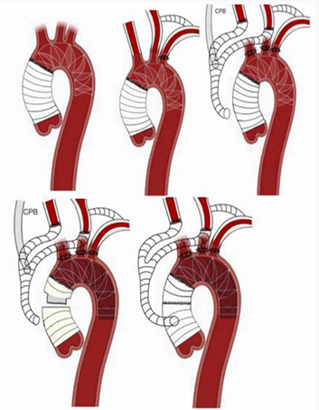
Figure 1: 1A: AMDS stent, Figure 1B: Left carotid-subclavian bypass, Figure 1C: Hybrid debranching, Figure 1D: Cook endoprosthesis, Figure 1E: Final result following reanastomosis
Patient recovered well without postoperative complications and was discharge home on day 9. At last follow-up, patient is well and without further complication or surgery.
DISCUSSION
The AMDS seems to offer effective initial result by promoting realignment of the intima, facilitating true lumen expansion, and ultimately reducing malperfusion [3]. However, the long-term durability of this prosthesis remains uncertain as some studies noted aortic diameter evolution [1]. Out of the 46 patients in Sabin J. et al study, 13% had disease related reintervention such as visceral stents, supra aortic vessel stents, redo Bentall procedures and more [3]. Multiple factors may contribute to this late progression of the aortic dissection, including distal stent-induced new entry (dSINE), excessive radial force exerted by the prosthesis, or persisting distal intimal tear that maintains FL pressurization [3]. Al Obaidi et al. reported two cases of aortic dilatation a few years following AMDS implantation that were managed using redo surgery using a minimally invasive frozen elephant trunk (FET) technique, deploying an E-vita OPEN NEO tri-furcated graft in zone 0 [3]. Their approach and ours share the same core principle which is avoiding complete AMDS explantation while excluding the persistent tear between true and false lumen with a covered stent graft. In their report, arch reconstruction was achieved using an integrated trifurcated FET prosthesis, which combines total arch replacement, supra-aortic vessel revascularization, and distal stent-graft deployment in one device. Antegrade trilateral cerebral perfusion was achieved using a balloon catheter. In contrast, our approach used a hybrid strategy, with supra-aortic debranching followed by antegrade deployment of a Cook covered endoprosthesis. This strategy allowed cerebral perfusion to be established independently of the systemic cooling via the Y graft connected to the innominate artery and left carotid. Therefore, isolating the brain from any emboli before going on systemic bypass.
CONCLUSION
This case highlights a strategic approach to managing post-AMDS dilatation. By combining a hybrid debranching with deployment of a zone 0 hybrid FET, the false lumen and DANE are excluded from systemic circulation. As late aortic dilatation appears to be a recurring long-term complication following AMDS placement, this method may become increasingly relevant in similar clinical scenarios. Our strategy illustrates a safe and reproducible option for AMDS reoperation by excluding DANE and arch vessels. It is not an option for suspicion of AMDS infection.
How to Cite
References
URL: https://pubmed.ncbi.nlm.nih.gov/38731123/
URL: https://www.sciencedirect.com/science/article/pii/S0022522322009412
URL: https://pubmed.ncbi.nlm.nih.gov/39402587/